You may also like...
Arsenal Roars to Premier League Glory, Parade Preparations Underway!

Former Vice President Atiku Abubakar congratulated Arsenal on winning the English Premier League, drawing parallels betw...
Scream Queen Jenna Ortega Teams Up With Visionary Director Leos Carax in Exclusive New Film!

Jenna Ortega will star in Leos Carax's next film, “Lily May B,” which was unveiled at Cannes and is set to begin shootin...
Iconic Japanese Franchise Returns: $80 Billion Behemoth Gets Live-Action Reboot!
The iconic Japanese franchise Hello Kitty is heading to Hollywood with a live-action/animation hybrid movie set to relea...
African Superstars Dominate BET Awards: Wizkid, Burna Boy, Asake, Tems Score Major Nominations

Nigerian music and the Afrobeats genre achieve significant global recognition at the 2026 BET Awards, with Wizkid, Burna...
Wizkid Makes History: First African Artist to Shatter 11 Billion Spotify Streams
Nigerian Afrobeats sensation Wizkid has set a new record, becoming the first African artist to achieve 11 billion stream...
Producer Unveils 'Entire Universes' for Characters in 'Margo's Got Money Troubles' Season 2
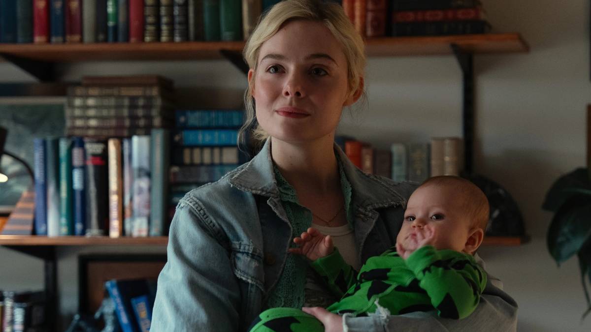
Collider's interview with producer Eva Anderson unveils key differences between <em>Margo's Got Money Troubles</em> show...
Uganda Unleashes Tourism Diplomacy to Entice Aussies
An Australian delegation's recent tour of Uganda concluded with strategic engagements aimed at boosting tourist arrivals...
Talk to Your Inbox: Google IO 2026 Reveals Revolutionary Gmail AI Integration
Google is enhancing Gmail with new conversational AI features, dubbed "Gmail Live," unveiled at the IO 2026 conference. ...